5
Thermo Scientific Poster Note
•
PN70513_E_04/14S
Conclusion
The GlycanPac AXH-1 column
charge, size, and polarity not p
LC-ESI-FTMS or FT-MS/MS a
and antibodies were carried ou
The GlycanPac AXH-1 column
separation and easy quantifica
The GlycanPac AXH-1 column
These new columns have high
stability.
The GlycanPac AXH-1 column
from proteins and mucins.
The GlycanPac AXH-1 column
glycosylaminoglycans and glyc
References
1. Varki, A. Biological Roles of Oli
Glycobiology
1993
,
3
, 97
–
130.
2. Bertozzi, C.R.; Freeze, H.H.; V
Pharmaceutical Industry
,
Esse
Harbor Laboratory Press: New
3. Guidance for Industry, Scientifi
Reference Product, Draft Guid
Food and Drug Administration,
GuidanceComplianceRegulato
Jan. 18, 2013).
4. Bigge, J.C. et al., Non-Selectiv
2-Amino Benzamide and Anthr
5. Apte, A; Meitei, N.S. Bioinform
Spectrometric Data Using Sim
Acknowledgeme
We would like to thank Mark Trac
from Thermo Fisher Scientific for
instruments.
from Proteins
h-performance LC/MS separation
r proteins. Analyzing unlabeled
nd cumbersome cleanup methods
rofile without adding further
5 shows the LC/MS analysis of
Pac AXH-1 column (1.9 µm). The
ze, and polarity. Using an
patible with MS detection, the
entation data for accurate
tographic peak. Native glycan
fluorescently labeled glycans,
escently labeled glycans generally
ks. The GlycanPac AXH-1 column
N
-glycans, depending on the
mple is not extremely limited,
AXH-1 is highly feasible.
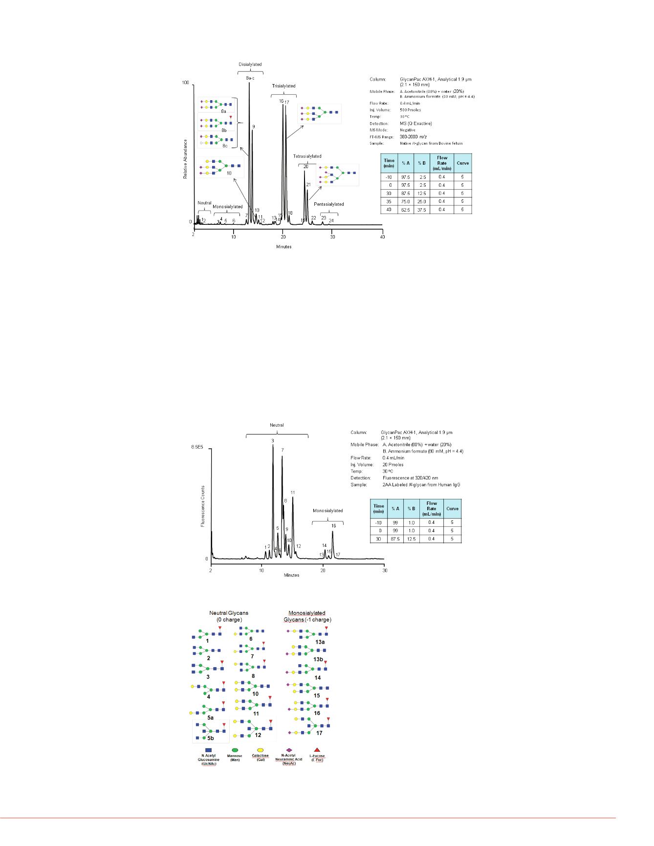
cans from Bovine fetuin by a
S detection.
cans from Bovine fetuin by the
ction.
FIGURE 5. LC-MS analysis of native
N
-glycans from Bovine fetuin. All the peaks are
detected by MS detection in negative ion mode.
Quantitative Determination of
Quantitative analysis of each gly
in protein batch comparisons an
profiles. In addition, quantitative
provide a tool for calculating the
enzymatic digestion with silidase
analysis of 2AB labeled
N
-glyca
(1.9 µm) with fluorescence detec
was estimated using a standard
the chromatographic analysis of
different amount of samples start
FIGURE 8: Quantitative estima
N
-glycan from Fetuin
Structural Analysis of
N
-Glycans from Antibodies by LC-MS Using
GlycanPac AXH-1 Column
Antibody research has gained significant interest as a part of the development of
protein biotherapeutics. Glycosylation of antibodies is a prime source of product
heterogeneity with respect to both structure and function. Variation in glycosylation is
one of the main factors in product batch-to-batch variation,
2-3
affecting product stability
in vivo
and significantly influencing Fc effector functions
in vivo.
A representative
example of the chromatographic separation of antibody glycans is shown in Figure 6,
where 2AA labeled
N
-glycans from IgG were separated using the GlycanPac AXH-1
column (1.9 µm). Characterization of glycans in each peak was performed by
LC-MS/MS and results are shown in Figure 7. Three different glycan charge states
were found in this human IgG; the majority of glycans are neutral or monosialylated,
with minor amounts of disialylated glycans. Separation of glycans based on charge
provides advantages compared to other commercially available HILIC columns.
FIGURE 6. Analysis of 2AA labeled
N
-glycans from human IgG.
FIGURE 7. Mass spectroscopic characterization of glycans in each Figure 6 peak.
registered trademark of PREMIER Biosoft
r Scientific and its subsidiaries.
in any manners that might infringe the intellectual
PO70513_E 1/13S


