4
Novel Glycan Column Technology for the LC-MS Analysis of Labeled and Native
N
-Glycans Released from Proteins and Antibodies
Charge, Size, and Polarity
for qualitative, quantitative, and structural
rged (neutral) and charged glycans present
the GlycanPac AXH-1 (1.9
μ
m, 2.1
×
150 mm)
paration and elution of glycans are based on
ed by the separation of acidic 2AB labeled
trisialylated, tetrasialylated and finally
arge state are further separated based on
each glycan charge state was confirmed
wn in Figure 2). Separation of glycans is
rovides significant structural and quantitative
own in Figures 1
and 2, detected by
formation about the separation of
N
-glycans
.
k was determined from the LC-MS study
n as shown in the following section.
LC-MS Analysis of Native Glycans Released from Proteins
The GlycanPac AXH-1 column is well suited for high-performance LC/MS separation
and analysis of native glycans from MAbs and other proteins. Analyzing unlabeled
glycans not only eliminates the extra reaction step and cumbersome cleanup methods
during labeling, but also retains the original glycan profile without adding further
ambiguity imposed by the labeling reaction. Figure 5 shows the LC/MS analysis of
native
N
-glycans from Bovine fetuin using the GlycaPac AXH-1 column (1.9 µm). The
native glycans were separated based on charge, size, and polarity. Using an
ammonium formate/acetonitrile gradient highly compatible with MS detection, the
separation enables excellent MS and MS/MS fragmentation data for accurate
confirmation of the glycan structure of each chromatographic peak. Native glycan
profiles are significantly different from the profile of fluorescently labeled glycans,
especially higher sialic acid glycans. However, fluorescently labeled glycans generally
provide better and more MS/MS fragmentation peaks. The GlycanPac AXH-1 column
is useful for the analysis of both native and labeled
N
-glycans, depending on the
amount of sample available. If the amount of the sample is not extremely limited,
analysis of unlabeled glycans using the GlycanPac AXH-1 is highly feasible.
glycans from Bovine fetuin by charge, size
-glycans standards and 2AB-
N
-glycans
beled
N
-Glycan Using GlycanPac AXH-1
n to MS was also explored. This is particularly
ructural information, enables in
-depth analysis
from bovine fetuin were separated on the
a Q Exactive mass spectrometer. Data-
all precursor ions (z< 2) and SimGlycan
idation. A representative example of the
tructural information obtained from the MS/MS
nPac AXH-1 column to separate glycans
r, coelution of different charge state glycans
ly available HILIC columns.
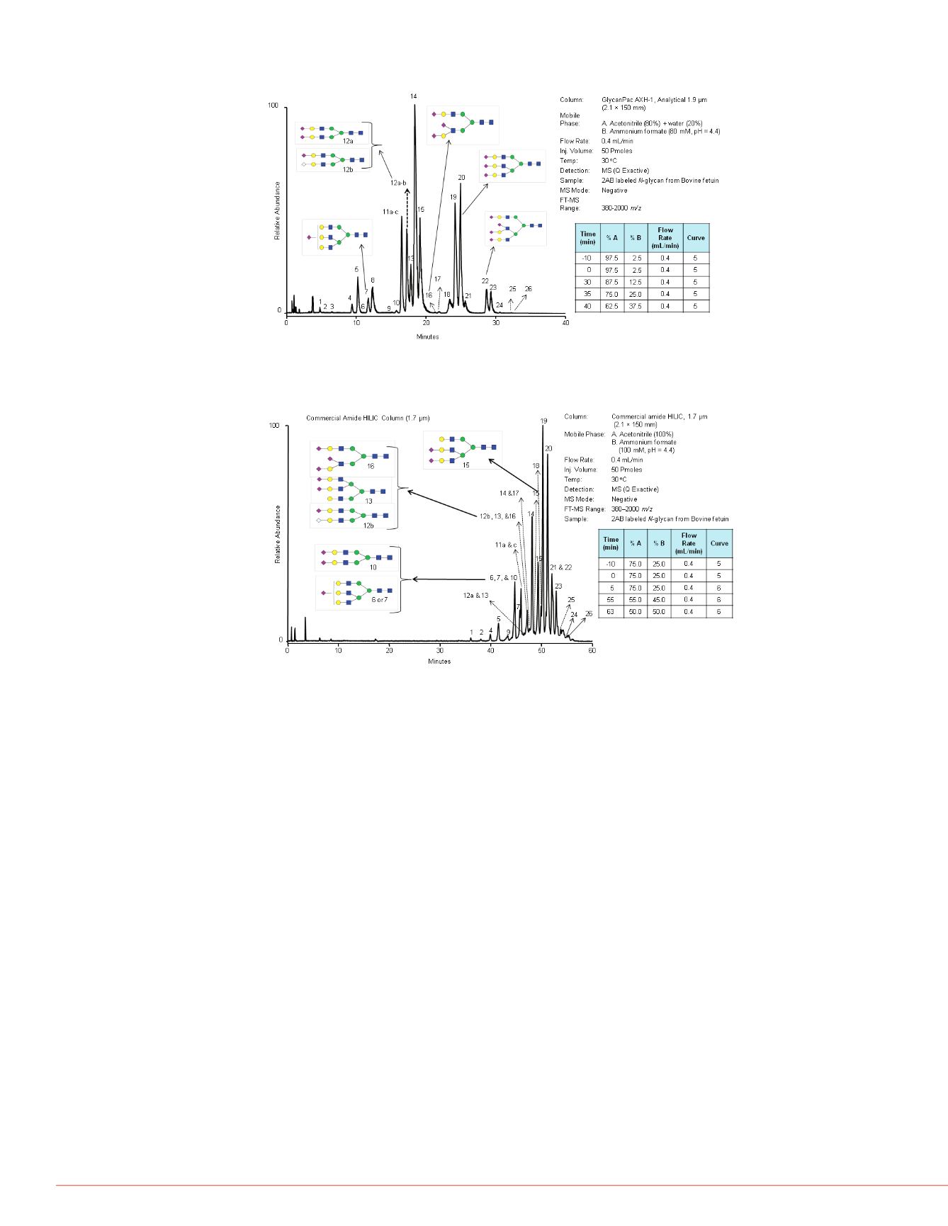
FIGURE 4. LC-MS analysis of 2AB labeled
N
-glycans from Bovine fetuin by a
commercial amide HILIC column (1.7 µm) with MS detection.
FIGURE 3. LC-MS analysis of 2AB labeled
N
-glycans from Bovine fetuin by the
GlycanPac AXH-1 (1.9 µm) column with MS detection.
FIGURE 5. LC-MS analysis of nati
detected by MS detection in negat
Structural Analysis of
N
-Glycan
GlycanPac AXH-1 Column
Antibody research has gained signifi
protein biotherapeutics. Glycosylatio
heterogeneity with respect to both st
one of the main factors in product ba
in vivo
and significantly influencing F
example of the chromatographic sep
where 2AA labeled
N
-glycans from I
column (1.9 µm). Characterization of
LC-MS/MS and results are shown in
were found in this human IgG; the m
with minor amounts of disialylated gl
provides advantages compared to ot
FIGURE 6. Analysis of 2AA labele
FIGURE 7. Mass spectroscopic ch
Prozyme is a registered trademark of ProZyme, Inc. SimGlycan is a registered trademark of PREMIER Biosoft
International. All other trademarks are the property of Thermo Fisher Scientific and its subsidiaries.
This information is not intended to encourage use of these products in any manners that might infringe the intellectual
property rights of others.
PO70513_E 1/13S


