
5
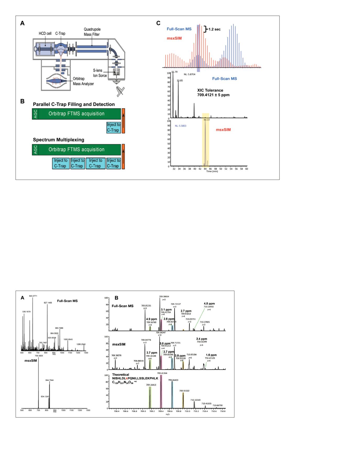
Figure 4. The Q Exactive mass spectrometer (A) is capable of multiple C-trap filling during Orbitrap detection (B) and producing a
multiplexed spectrum (C).
Multiplexing the SIM event facilitated selective data
acquisition for two additional active-site peptides that
co-eluted in the same retention time window as the ULK3
active-site peptide (Figure 5A). Despite a large number of
background ions in the full-scan MS scan, three target
m/z
values are easily acquired in the msxSIM window.
Quadrupole mass filtering around the targeted
m/z
values
enables greater accumulation of the target peptide precursor
ions. Figure 5B shows the narrow mass region centered at
the
NISHLDL
K
PQNILLSSLEKPHLK
+4 precursor
m/z
value for the full-scan MS, SIM, and theoretical isotopic
distribution. Both full MS and SIM scans show a co-isolated
matrix ion in the +3 charge state. However, the high
resolution of the Orbitrap mass analyzer facilitates
separation of the background signal from the targeted
peptide even though the mass difference between the
matrix ion and the A+2 target isotope is only 0.05 Da
(76 ppm) at 20% relative intensity. The SIM event also
provided greater fine structure compared to full-scan MS
with increased signals for target peptide isotopes. Each
peptide was confirmed in a separate targeted MS
2
experiment. Excellent retention-time correlation was
observed between targeted msxSIM and targeted MS
2
experiments (data not shown).
Figure 5. Comparative HR/AM mass spectra for the targeted active-site peptide
NISHLDL
K
PQNILLSSLEKPHLK
between full-scan MS and msxSIM at a determined retention time (A). Zoom in shows the +4 precursor charge
state compared to the theoretical precursor isotopic distribution used for verification (B).


