5
Thermo Scientific Poster Note
•
PN-64155-ASMS-EN-0614S
Conclusion
Two different digestion way
while the other used the co
10 out of 16 disulfide bonds
and 13 out of 16 were foun
complementary.
Totally, 15 out of 16 disulfid
MS/MS information.
Biopharmaceutical industry
Orbitrap Elite, with reprodu
References
1. Nebija D., Kopelent-Frank
Comparison of two-dimensi
MS analysis of therapeutic
rituximab.
Journal of Pharm
2. Martin Samonig, Christian
Monoclonal Antibody Rituxi
Spectrometer.
3.
FIGURE 8. All of the identified d
bond can be found in both exp
found in trypsin-only digestion.
only in double digestion.
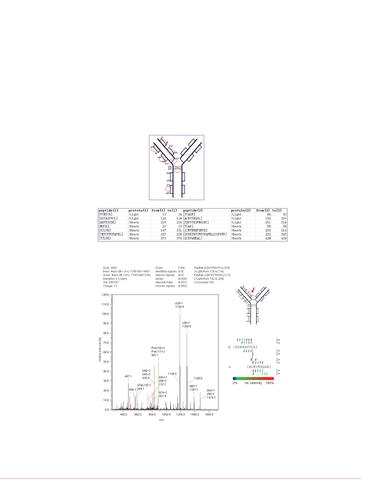
FIGURE 7. The annotated MS/MS spectrum of disulfide bond C133-C193 on light
chain (double digestion).
the trypsin-only digested
d disulfide bonds.
FIGURE 7 shows the disulfide bon
this double digestion experiment,
time. Because the disulfide bond-in
shorter peptides, double digestion i
In double digestion result, we can f
heavy chain and one of the interch
weren’t identified in
trypsin only ex
linkage on heavy chain was found i
double digestion condition; this can
linkage was cut into some very sho
beyond the detection line of mass
By combination of these two result
identified in our experiment succes
d in two different ways.
ypsin-only digested
fide bond C265-C325 on
FIGURE 5 is an annotated MS/MS spectrum of disulfide linkage between Cys265 and
Cys325 on heavy chain. The accuracy of precursor ion is high (2.19ppm), which
indicated that an Orbitrap MS can produce high mass accurate data. It’s also easily to
find that many continuous, disulfide linkage-included fragment ions were identified.
These ions are strong evidence which suggest the existence of the disulfide bond.
The result of double digested sample
In our experiments, we also tried double digestion, to produce more disulfide linkage-
included peptides which were suitable for mass spectrometer detection. Chymotrypsin
and trypsin were used for double digestion, which can get nearly 100% sequence
coverage of the sample (FIGURE 3).
By using the software StavroX
TM
, we have identified 13 disulfide bonds in the double
digested Rituximab. FIGURE 6 shows the summary of all identified disulfide bonds in
double digested sample.
FIGURE 6. All of the identified disulfide linkage in double digested Rituximab.
Rituxin is a registered trademark of Biogen I
Ltd.
StavroX is a trademark of Martin-Luther Uni
property of Thermo Fisher Scientific and its
This information is not intended to encourag
intellectual property rights of others.


