36
For more information, or to download product instructions,
Thermo Scientific
Ninhydrin Detection Reagents for Amino Acids
Ninhydrin-based monitoring systems are among the most
widely used methods for quantitatively determining amino
acids after they are separated by ion exchange
chromatography.
The color reaction between amino-containing compounds
and ninhydrin (2,2-dihydroxy-1,3-indandione) is very
sensitive. McCaldin has studied all phases of ninhydrin
chemistry and proposed a mechanism for the reaction of
ninhydrin with amino acids, accounting for the aldehydes,
carbon dioxide, ammonia and hydrindantin known to be
produced.
1
A yellow colored product (monitored at 440 nm)
is formed upon reaction with the secondary amino acids,
proline and hydroxyproline.
2
Ninhydrin decarboxylates and
deaminates the primary amino acids, forming the purple
complex known as Ruhemann’s Purple,
3
which absorbs
maximally at 570 nm.
Ninhydrin chemistry was adapted to a fully automatic,
two-column amino acid analysis procedure in 1958 by
Spackman, Stein and Moore.
4
Moore and Stein defined the
requirements for a reducing agent (such as stannous
chloride) to achieve reproducible color values for amino
acids monitored with ninhydrin.
5
Titanous chloride was
reported by James to eliminate precipitates encountered
when using stannous chloride.
6-8
Methyl Cellosolve
®
(ethylene glycol monomethyl ether) buffered with 4 M
sodium acetate at pH 5.51,
9
and dimethylsulfoxide (DMSO)
buffered with 4 M lithium acetate at pH 5.20
10
are the most
common solvents used for ninhydrin. DMSO remains stable
longer than Methyl Cellosolve, particularly when kept
chilled. These ninhydrin reagent solutions, with increased
stability, were also reported by Kirschenbaum.
11
Sensitivity of the ninhydrin system depends on several
factors. Amino acids produce slightly different color yields,
and these values may vary from one reagent preparation
to the next. Ninhydrin also is sensitive to light, atmospheric
oxygen and changes in pH and temperature. When
ninhydrin becomes oxidized, its color does not develop well
at 570 nm, but absorption at 440 nm remains fairly constant.
When the height of the proline peak at 440 nm approaches
the height of the glutamic acid peak at 570 nm, for equal
amounts of each, reagent degradation is indicated.
References
1. McCaldin, D.J. (1960).
Chem. Rev.
60
, 39.
2. Hamilton, P.B. (1966).
Advan. Chromatogr.
2
, 3.
3. Ruhemann, J. (1911).
J. Chem. Soc. London
99
, 797.
4. Spackman, D.H., Stein, W.H. and Moore, S. (1958).
Anal. Chem.
30
, 1190.
5. Moore, S. and Stein, W.H. (1954).
J. Biol. Chem.
211
, 907.
6. James, L.B. (1971).
J. Chromatogr.
59
, 178.
7. James, L.B. (1978).
J. Chromatogr.
152
, 298-300.
8. James, L.B. (1984).
J. Chromatogr.
284
, 97-103.
9. Moore, S. and Stein, W.H. (1948).
J. Biol. Chem.
176
, 367.
10. Moore, S. (1968).
J. Biol. Chem.
243
, 6281-6283.
11. Kirschenbaum, D.M. (1965).
Anal. Biochem.
12
, 189.
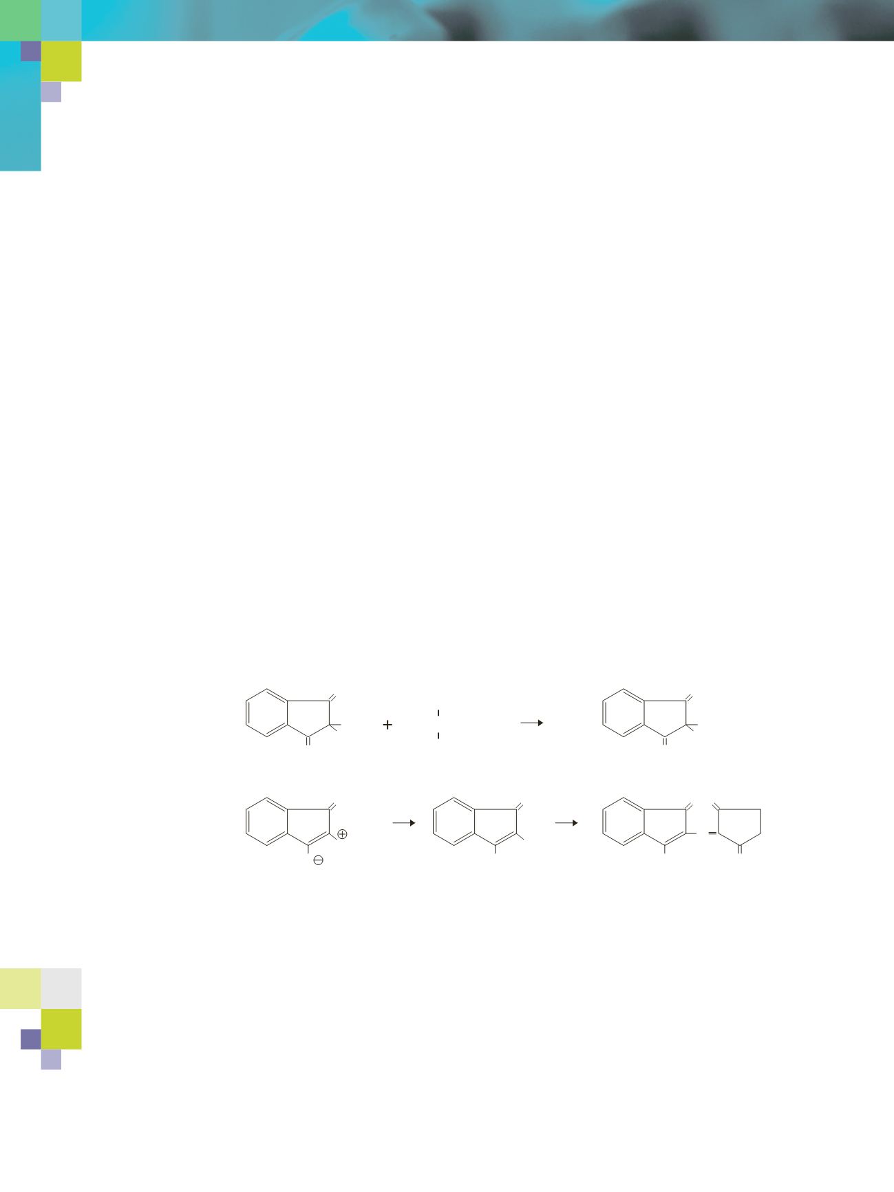
Reaction Scheme.
The course of the ninhydrin reaction with amino acids is as follows:
1. Ninhydrin (2,2-dihydroxy-1,3-indandione) reacted with amino acid.
2. The intermediate formed as the first reaction product.
3. Intermediate gives rise to dipolar ion by decarboxylation and dehydration.
4. The dipolar ion hydrolyzes, producing the amine.
5. The amine condenses with a second molecule of ninhydrin to give Ruhemann’s Purple.
OH
O
OH
O
(1)
–H
2
0
COOH
R–C–NH
2
H
OH
O
NH–CHR
•
COOH
O
(2)
O
NH=CHR
O
(3)
O
NH
2
O
(4)
O O
N
OH
O
(5) Ruhemann's Purple


