
2
Enrichment of EGFR/PI3K/AKT/PTEN Proteins for Research using Immunoprecipitation and with Mass Spectrometry-based Analysis
Overview
Purpose:
Identification and quantification of EGFR/PI3K/AKT/PTEN proteins for
research using an optimized immunoprecipitation to mass spectrometry (IP-MS)
workflow.
Methods:
We evaluated immunoprecipitation with directly coupled antibodies or
biotinylated antibodies with immobilized streptavidin resin. EGFR, PI3K, AKT isoforms
and PTEN were enriched from two cell lysates using an optimized IP to MS workflow. A
multiplex, targeted selected reaction monitoring (SRM)-based MS research method
was developed to measure the limit of quantitation (LLOQ) of EGFR, AKT2, AKT1,
PTEN, PIK3CA and PIK3R1 tryptic peptides. Multiple targets (EGFR, AKT isoforms,
PTEN) were immunoprecipitated simultaneously and quantified by targeted SRM
assay.
Results:
Immunoprecipitation using magnetic beads resulted in overall higher yield of
target protein and less non-specific binding than agarose beads for MS research
applications. Enrichment of EGFR, AKT2, AKT1 and PTEN from two cell lysates
enabled MS detection and quantitation. Enrichment of as low as 7ng recombinant
EGFR in human plasma matrix allowed absolute quantitation by LC-SRM. Multiplexed
target immunoprecipitation resulted in simultaneous identification and quantitation by
MS.
Introduction
A major bottleneck in the verification of protein biomarkers in clinical research is the
lack of methods/reagents to quantify medium to low levels of proteins of interest in
human samples. Immunoprecipitation (IP) and mass spectrometry (MS) are
complementary techniques that permit sensitive and selective characterization and
quantitation of low abundance protein analytes in cell lines, tissue, and biofluids. IP
provides both enrichment and high selectivity while the MS provides high selectivity,
sensitivity, and multiplexing across a range of analyte concentrations in different
matrices. The quantitative evaluation of protein expression and PTM status of EGFR-
PI3K-AKT signaling pathway proteins enables the precise characterization of the
disease.
Methods
Sample Preparation
EGFR from A431 lysate was immunoprecipitated by direct IP methods (Hydrazide
activated polyacrylamide bead, Aldehyde activated agarose bead, NHS-Ester activated
magnetic bead, and Epoxy activated magnetic bead) and indirect IP methods (Streptavidin
coated polyacrylamide
bead and Thermo Scientific™ Pierce™
Streptavidin magnetic
bead). IP eluted samples were evaluated by western blot and in-solution trypsin digestion
followed by MS analysis. IP conditions were optimized for enrichment of medium to low
abundant targets (EGFR, AKT isoforms, PTEN and PIK3CA) for MS applications. Multiplex
IP was performed to enrich EGFR, AKT isoforms and PTEN targets simultaneously from
HEK293 lysate with biotinylated antibodies and with Pierce Streptavidin coated magnetic
beads.
Liquid Chromatography and Mass Spectrometry
IP eluates were reconstituted in 6M Urea, 50mM Tris-HCl pH 8 followed by reduction,
alkylation and trypsin digestion overnight. Prior to MS analysis, tryptic digest samples were
desalted using the Thermo Scientific™ Pierce™ C18 Spin Tips. For discovery MS, the
samples were analyzed by LC-MS/MS using a nanoLC system at 300 nL/min over a 45
min gradient and Thermo Scientific™
Orbitrap
XL ™ mass spectrometer (DDA, Top 6,
CID). For targeted MS, the samples were analyzed by LC-SRM/MS with the Thermo
Scientific ™ TSQ Vantage ™ mass spectrometer and Thermo Scientific™ Easy
nanoLC II
system.
Data Analysis
Discovery MS data were analyzed with Thermo Scientific™ Proteome Discoverer™ 1.4
and Scaffold 4.0 software to assess percent sequence coverage, spectral counts and
PTMs. For targeted LC-
SRM/MS data analysis, Thermo Scientific ™ Pinpoint™ and
Skyline
software
were used to measure limit of quantitation (LOQ ) and target analyte
concentration.
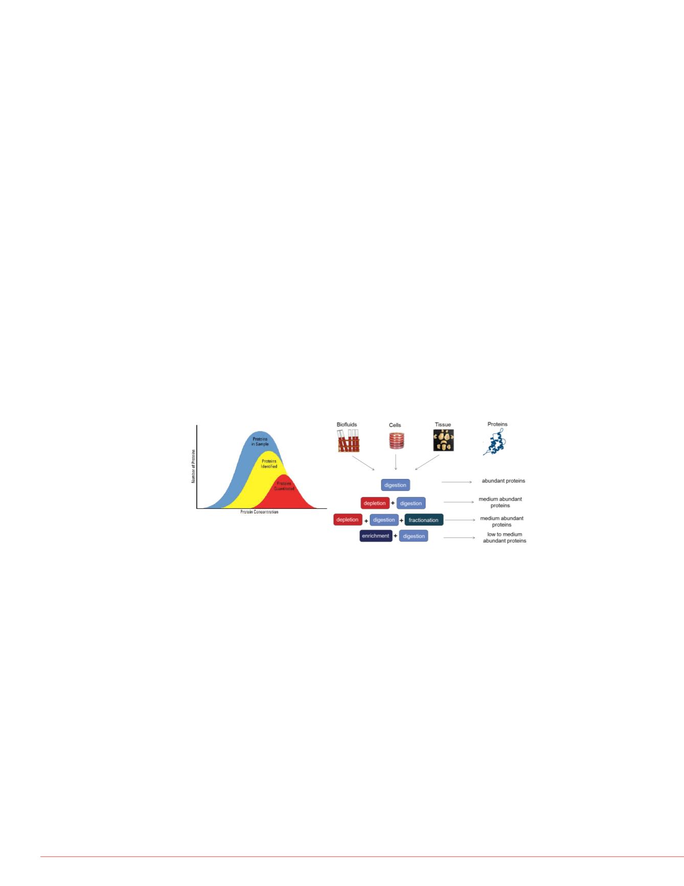
FIGURE 1. Enrichment is necessary for medium to low abundant proteins.
Results
Benefits of Magnetic Beads
•
Lower background: Minimal
•
High signal to noise: Easy a
chance of losing sample
•
Easy handling: Easy separa
•
Time and effort: Less washi
•
Better reproducibility: Produ
•
Ab savings: All binding on o
•
Automation: Improves throu
FIGURE 3. Evaluation of EG
EGFR immunoprecipitation w
biotinylated antibody with im
determined by Western blot.
were determined by LC-MS/
beads resulted in fewer back
coverage.
FIGURE 2. Experimental wo
Protein targets are immune-e
Western blot after gel electro
analyzed by nLC-MS/MS to i
labeled, quantitative peptide
research methods for absolut
Anti-EGFR Western
P: Polyacrylamide, A: Ag
M: Magnetic
+ Anti-EGFR; - Rabbit


